Why Primary Cell Culture Requires a Different Approach
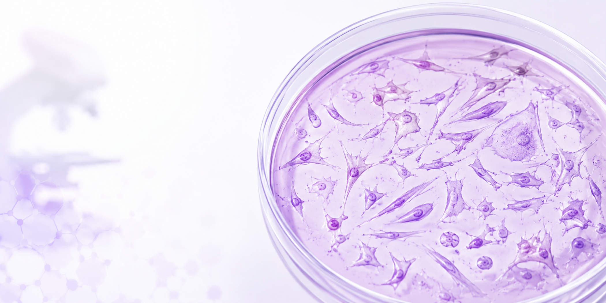
Unlike immortalized cell lines, primary cells are directly isolated from tissue and still retain much of their native biology. This makes them highly valuable for studying physiology, drug response, differentiation, and disease models. However, it also makes them significantly more fragile, variable, and difficult to maintain.
Many beginners approach primary cell culture using workflows learned from robust immortalized lines, only to find that the cells attach poorly, differentiate unexpectedly, or die within days.
The reality is that primary cells require a different mindset. Success often depends less on aggressive manipulation and more on minimizing stress and preserving the biological state of the cells.
Work Fast: Tissue Quality Declines Quickly
Primary cell culture quality starts before the cells are even plated. Fresh tissue is commonly stored in a preservation buffer or transport medium to slow degradation and maintain viability during transport. However, once tissue is removed from the organism, the clock starts ticking.
Cell viability decreases rapidly over time. For many tissues, processing within approximately 4 hours is ideal. The longer the delay, the greater the risk of:
cellular stress
hypoxia
apoptosis
necrosis
loss of attachment potential
This does not mean rushing carelessly, but rather preparing all reagents, media, coatings, and tools beforehand so the isolation workflow can proceed efficiently.
Freezing tissue before isolation can also dramatically reduce viability and recovery, especially for fragile primary populations. While cryopreservation protocols exist for some tissues, freshly processed samples generally provide better attachment and survival.
Leave the Cells Alone
One of the most common beginner mistakes is disturbing primary cells too frequently after plating.
With immortalized cell lines, routine handling is often tolerated. Primary cells are different.
After initial seeding:
avoid excessive movement
avoid repeated media changes
avoid constant microscopy checks
avoid unnecessary temperature fluctuations
Freshly isolated cells are attempting to attach, recover from dissociation stress, re-establish signaling, and adapt to artificial culture conditions. Too much disturbance during this stage can reduce attachment efficiency and survival. One of the biggest mindset shifts for beginners is realizing that primary culture is not simply about maximizing growth. It is about preserving biology. Every manipulation, including: media change, dissociation, centrifugation, temperature fluctuation, over-confluency, and nutrient depletion can influence cellular state.
Primary cells often behave more like living tissue than standardized laboratory reagents. The less unnecessary stress introduced into the workflow, the better the chances of maintaining biologically meaningful culture.
In many cases, less intervention leads to better outcomes. Minimal feeding during the first few days is often beneficial unless the protocol specifically requires otherwise.
Use Mild Trypsin Conditions
Primary cells are usually far more sensitive to enzymatic dissociation than immortalized lines. Harsh trypsinization can:
damage membranes
cleave surface proteins
alter receptor expression
reduce viability
increase differentiation stress
Whenever possible:
use lower trypsin concentrations
shorten exposure times
neutralize quickly
monitor detachment carefully
Over-trypsinization is one of the fastest ways to lose fragile primary cultures. Some cell types may also benefit from gentler dissociation reagents or enzyme-free approaches.
Use High FBS Early, Then Gradually Reduce It
During the initial establishment phase, primary cells often require higher serum concentrations to maximize survival and recovery. Higher FBS concentrations provide:
growth factors
attachment support
nutrients
stress protection
For many workflows, elevated serum conditions are maintained during the first 1–2 weeks while the culture stabilizes. Afterward, serum concentration is gradually reduced depending on:
the cell type
experimental goals
differentiation state
downstream applications
Abrupt serum reduction can shock fragile cultures, so gradual adaptation is generally preferred.
Coating Matters More Than You Think
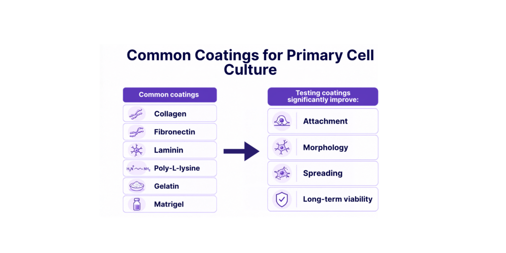
Different primary cells prefer different extracellular environments. A coating that works perfectly for one cell type may fail completely for another.
Morphology Is One of Your Most Important Readouts
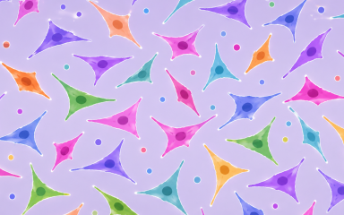
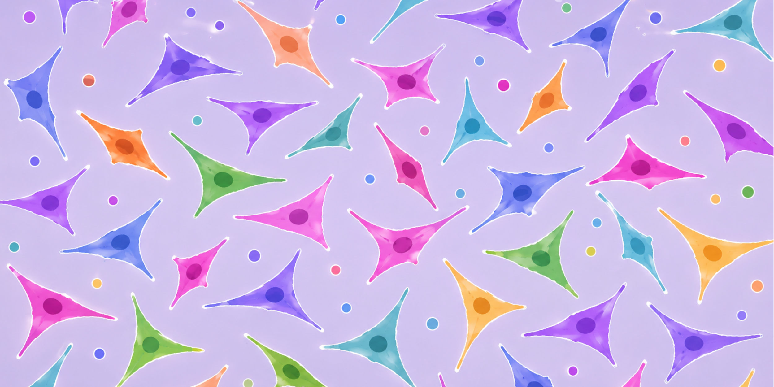
Primary cell cultures are highly heterogeneous. Morphology often provides the first indication of:
stress
differentiation
contamination
senescence
attachment problems
Researchers frequently use staining methods to identify and differentiate cell populations within mixed cultures. Depending on the application, staining may be used to evaluate, lineage markers, differentiation markers, structural proteins, viability, and etc.
Monitoring morphology consistently over time is essential for understanding whether the culture is stabilizing or drifting biologically.
It raises an interesting question: what if one day we could distinguish different cell populations directly from microscopy images without fully dissociating and sorting the cells?
As AI image analysis continues to improve, morphology, texture, and spatial patterns may help identify heterogeneous cell populations directly within intact primary cultures. This could function almost like sorting cells through an image while preserving their biological context.
Use Imaging to Monitor Growth Without Disturbing the Culture
Many decisions in primary cell culture are still based on visual inspection. Researchers assess whether cells have attached, whether the culture is expanding, and whether morphology is changing. These assessments are useful, but they can also be subjective and user-dependent.
Image-based measurements can provide a more consistent and non-destructive way to monitor:
- monitoring attachment efficiency
- quantifying confluency
- measuring morphology
- tracking differentiation
- detecting abnormal populations
Adherent cell count and confluency provide different information. Cell count estimates how many cells are present, while confluency measures how much of the culture surface is covered.
For example:
- 5,000 small, compact cells may occupy less surface area than 1,000 large, spread cells
- Stressed cells may remain attached but change morphology significantly
- Over-confluent cultures may alter their differentiation state before obvious cell death occurs
This is why measuring cell growth using both adherent cell count and confluency together can provide a much more complete understanding of culture health. Rather than relying only on visual estimation or total count alone, combining these measurements helps researchers better interpret:
- attachment efficiency
- proliferation
- spreading behavior
- culture readiness
- biological stability
This becomes especially important in primary cultures, where small environmental or handling changes can significantly affect cellular behavior. In primary culture, cell growth is more than proliferation alone. It is the combination of count, confluency, morphology, and attachment that reveals how healthy a culture truly is.
To Sum Up
Primary cell culture is often less forgiving than standard cell line work, but it also provides biology that is much closer to physiological reality.
For beginners, the most important lesson is usually not to manipulate cells more, but to disturb them less.
Work quickly. Handle gently. Optimize conditions carefully. Observe morphology closely.
In primary culture, small handling decisions can have major biological consequences.
Continue Exploring
Learn how confluency estimation introduces variability in routine cell culture:
Cell Confluency: When It Works—and When It Fails
For a practical workflow, see our guide to image-based adherent cell counting and learn how sensitive cultures can be monitored without detachment.
Cell Counting in Cell Culture: How to Reduce Variability – SnapCyte
Nader Al Nakouzi
I am a cancer biologist and translational researcher with experience in cell culture, microscopy, image-based assays, and therapeutic development. My work focuses on understanding complex biological systems and improving how experimental data is measured, interpreted, and translated into meaningful decisions.
Track Cell Growth From Images
Sign up free. Get 25 credits. No credit card required.