Summary
Art and science, although seemingly very different, often coexist and complement each other. In some cases, one can even help you understand the other. This is especially true for cellular biology. For those of us who work in the field, we can attest to how cellular biology can be both informative and beautiful. As cells grow, they create an intricate mosaic that gives you insights into their health, interactions with each other, and their response to the environment. Properly setting up experiments and ensuring result consistency is truly an art. Here, we discuss what visualizing cell confluency, morphology and patterns can tell you and how to use the visual appeal of cells to your advantage.
Understanding Cellular Art Signatures
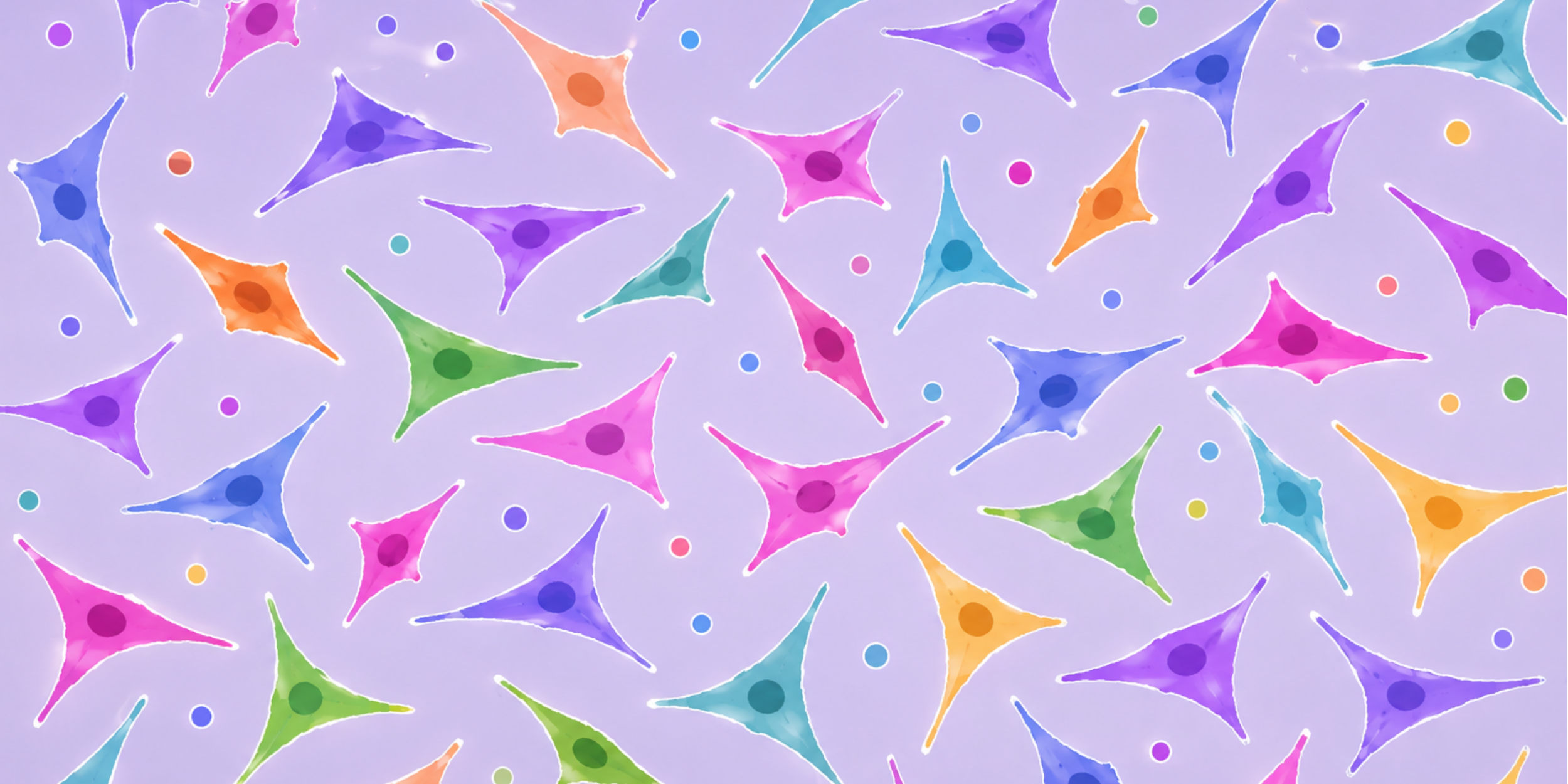
Every artist has their own identifiable style. Picasso is known for his use of interesting shapes, predominantly his use of cubes. Monet often painted pictures of his garden in light airy brush strokes. Different art mediums and brush sizes also have their own characteristics that generate a certain look. Similarly, individual cell lines have their own unique vibrancy and quirks that make them interesting.
Some cell lines form distinct patterns, some vary in the time it takes them to divide and fill the canvas space, and some are sensitive to their surroundings and what they grow on. The cell size, shape, and arrangement are all factors to carefully observe and take into consideration when culturing or performing experiments.
One of the major uses of these artistic signatures is to figure out timing. When you use different mediums or styles, it can affect how long it takes to complete your art piece. Oil paintings take days to dry before being able to add another layer, while acrylic paints dry within a few minutes. Hyper-realism pieces that use teeny, tiny strokes, will take significantly longer than if you were to paint a single circle on a plain background (yes, this is still considered art).
Metastatic cancer cells, for instance, are more like acrylics and painting a circle. They grow fast and are quick to take up space. Primary cell cultures, on the other hand, are like oil and hyper-realism pieces. They are slow and require meticulous care. Thus, whether you need to subculture or plate for an experiment, how quickly they take up space, and how fast they grow are all factors that affect how you take care of them and seed them for downstream experimental work.
As you continue to work with your cell lines of interest, you become familiar with their signature appearance and behavior. Any deviation from this signature could be indicative of changes intracellularly or as a response to the environment and it is important to investigate what these changes are trying to ‘tell’ you about the condition of your cells.
Check out the Everything You Need to Know About Cell Confluency post to learn what influences the confluency of your cell culture.
Expressive Cellular Expression
Often, changes in an art piece can be reflective of changes in the artist’s own inner feelings and mental well-being. Starry Night is one of Van Gogh’s most iconic paintings and is often admired for its beauty. Interestingly, the swirling nature of the brush strokes is often interpreted as a representation of his turbulent emotional state (1).
This concept of external appearance being a reflection of internal changes can also be applied to cell culture. This is not to say that cells have feelings (that we know of), but changes in cellular morphology and confluency can indicate changes internally at the genetic and protein expression level.
These changes in appearance can be a result of intrinsic or extrinsic forces. For example, if a cell line has been in culture for too long, it can begin to drift genetically from the original source and behave accordingly (2). Alternatively, changing environmental factors can cause differences in cell behavior and appearance, such as adding a drug that inhibits or suppresses the activity of certain proteins, which in turn slows growth and/or kills the cells.
The reverse is also true. Artists often use certain colors and tones in their pieces to convey a certain feeling. Darker colours project sadness and bright colours can invoke joy. The same way that art can change our feelings, cell confluency and cell distribution in their culture vessel can alter gene and protein expression. For instance, high density confluency can trigger adaptive changes to cells that increase the expression of certain proteins and pathways as a possible way to facilitate survival (3).
You can tell a lot from how cells look. These visual observations can provide valuable insight into understanding cells at the genetic and protein levels. Confluency and cellular morphology can also be a way of ensuring data consistency within cell biology.

Symmetry in Cell Culture: More Than Just Aesthetics
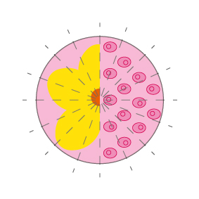
In art, symmetry serves as a sense of visual balance and harmony. In nature, flower petals are radially symmetrical to maximize sunlight intake optimal for attracting pollinators (4). Symmetry in 2D cell culture, like in art and nature, is not only aesthetically pleasing but actually serves a function.
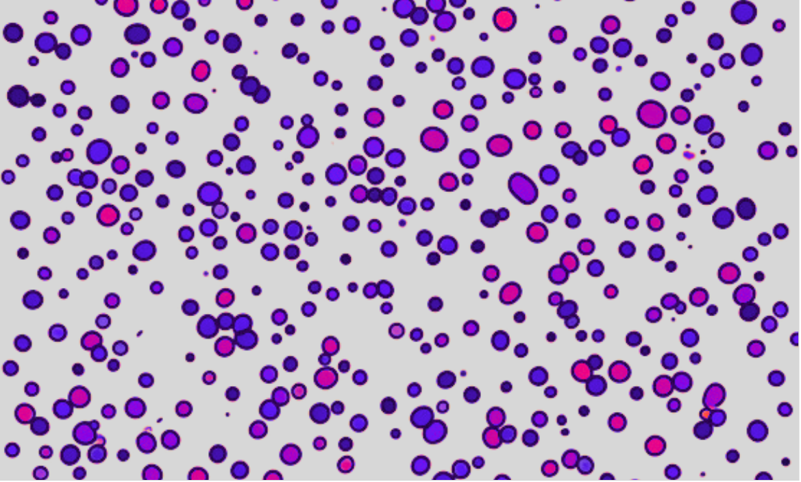
Asymmetry, in art on the other hand, is often tied with chaos (5). Some asymmetry is not necessarily a bad thing, however, the more asymmetry an art piece displays, the more unsettled the viewer can feel. Asymmetry in cell culture is not only visually unsatisfying but this cellular chaos can actually affect experimental outcomes. When cells aren’t seeded properly and there is variability in density, it can alter the response to drug treatments and result in inconsistencies in experimental outcomes (6).
Here we can see that artistic principles of symmetry are prevalent in cell culture. Elements of symmetry are not only concepts within art but serve valuable functions in cellular assays and cell maintenance. By following the rules of symmetry we can achieve cellular balance and harmony.
Visual Tips for Effective Subculturing and Seeding
Working with cells, you can analyze and interpret their visual appearance in order to optimize maintenance cultures and experimental outcomes. Here are some visual tips and tricks we use to make sure our cells are happy and healthy – we hope they work for you, too:
- Regularly check distribution of cells before and after passaging and seeding for an experiment.
- Look at different areas of the plate or well to ensure even distribution of cells after seeding a new plate or flask.
- For adherent cells, lots of floating cells when there is a minimal change in confluency over time may indicate crowding or overly dense sections.
- Cells that are slow growing and take longer than usual to reach a confluent state can indicate that cells are seeded too sparsely.
- Cells should be passaged if some sections are getting very confluent and other sections aren’t as confluent.
Download our eBook: How to Measure Cell Confluency comparing methods to measure confluency.
Conclusion
In biological research, visualizing cell confluency transcends more than just observation; it is an artform that merges scientific inquiry with aesthetic appreciation. As researchers continue to unravel the impact of cellular distribution and their growth environment, microscopy is a portal into a world where each image captured tells a unique story. Art, although seemingly different from science, has concepts within it that aid in understanding the importance of cell confluency. We hope you see that visualizing cell confluency is art and science that is a testament to the beauty and complexity of life at the cellular level.
References
- Museum of Modern Art (MoMA). Vincent van Gogh: Starry Night. Retrieved from https://www.moma.org/collection/works/79802
- Hughes P, Marshall D, Reid Y, Parkes H, Gelber C. The costs of using unauthenticated, over-passaged cell lines: how much more data do we need? Biotechniques. 2007 Nov;43(5):575, 577-8, 581-2 passim. doi: 10.2144/000112598. Erratum in: Biotechniques. 2008 Jan;44(1):47. PMID: 18072586.
- Trajkovic K, Valdez C, Ysselstein D, Krainc D. Fluctuations in cell density alter protein markers of multiple cellular compartments, confounding experimental outcomes. PLoS One. 2019 Feb 4;14(2):e0211727. doi: 10.1371/journal.pone.0211727. PMID: 30716115; PMCID: PMC6361456.
- Damerval, C., Jabbour, F., Nadot, S., Citerne, H.L. (2017). Evolution of Symmetry in Plants. In: Nuno de la Rosa, L., Müller, G. (eds) Evolutionary Developmental Biology. Springer, Cham. https://doi.org/10.1007/978-3-319-33038-9_59-1
- McMANUS IC. Symmetry and asymmetry in aesthetics and the arts. European Review. 2005;13(S2):157-180. doi:10.1017/S1062798705000736
- Xue Z, Zeng J, Li Y, Meng B, Gong X, Zhao Y, Dai X. Proteomics reveals that cell density could affect the efficacy of drug treatment. Biochem Biophys Rep. 2022 Dec 9;33:101403. doi: 10.1016/j.bbrep.2022.101403. PMID: 36561432; PMCID: PMC9763681.

")
")
")