AI Image Analysis for Microscopy

SnapCyte™ provides AI image analysis for microscopy, delivering fast, reproducible insights from 2D and 3D cell images.

What We Offer

Tailored AI Solutions for Complex Microscopy

Custom AI Model Development

We build deep learning segmentation models tailored to your samples. including complex 2D and 3D structures like spheroids, co-cultures, organoids, IHC-stained tissue, and other microscopy formats.

MLOps/Data Infrastructure

We deploy and maintain scalable pipelines to manage your models and imaging data. From training to inference, we ensure reproducibility and performance at scale.

Data Analysis Services

Our scientific team helps define analysis parameters and delivers expert quantification of microscopy data—no coding or in-house AI expertise required.

What Our AI Image Analysis Models Can Do

Custom AI Image Analysis for Microscopy

Our platform delivers AI image analysis for microscopy, tailored for both research and commercial workflows.

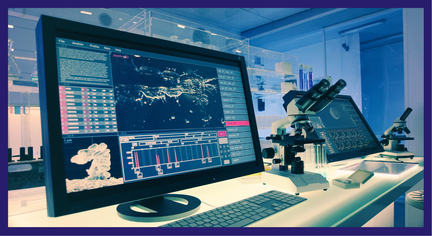
SnapCyte™ models enable precise quantification and analysis of key parameters in 2D microscopy images, including cell count, area, size, and morphology. Our solutions also measure intensity across both brightfield and fluorescence images, providing comprehensive insights into cell characteristics.

SnapCyte™ brings advanced capabilities to 3D microscopy, enabling detailed analysis of cell count, size, morphology, and area within volumetric datasets.

Track and analyze cell movement seamlessly with SnapCyte™ models. Our solutions capture individual cell trajectories, quantify cell migration, and provide dynamic insights into cell interactions and behavior over time.

Learn more

Deep learning cell segmentation count

Ready to Accelerate Your Image Analysis?

Contact us today to discuss your needs and discover how we can deliver tailored AI solutions.

What scientists say about us

News and resources

Latest insight and updates