Invasion Assay Data in less than 30 minutes with AI
“SnapCyte’s integration has significantly enhanced the precision and efficiency of our invasion assays, providing a solid answer to our previous data analysis challenge. We’ve shifted from spending hours on taking the images and figuring out various parameters on other platforms to analyzing our data within 30 minutes.”
Dr. Eliana Beraldi, Senior Research Assistant The Somasekharan Lab, Vancouver Prostate Centre.
Automating Transwell Invasion Assay with AI-Based Image Analysis
The Vancouver Prostate Centre is a leading entity in prostate cancer research, committed to elucidating cancer progression’s mechanisms and pioneering therapeutic innovations. Dr. Syam Somasekharan, a prominent research scientist at the Centre, spearheads pivotal studies on RNA regulatory networks, focusing on stress adaptation and treatment resistance in prostate cancer.
Challenges
In their work on prostate cancer research, Dr. Somasekharan’s team faced challenges in capturing usable microscope images for analysis, documentation and publication. Furthermore, while exploring how to analyze their experimental data, they found that image processing software required extensive time and training to use.
The team also evaluated traditional methods for measuring cell invasion based on reading the optical density of dissolved stains but were discouraged by several drawbacks. These included the time-consuming nature of traditional methods (about an hour per plate), the necessity for training and experience, the irreversible sample loss and the inability to visually verify data.
Optimal Assay Analysis with SnapCyte™
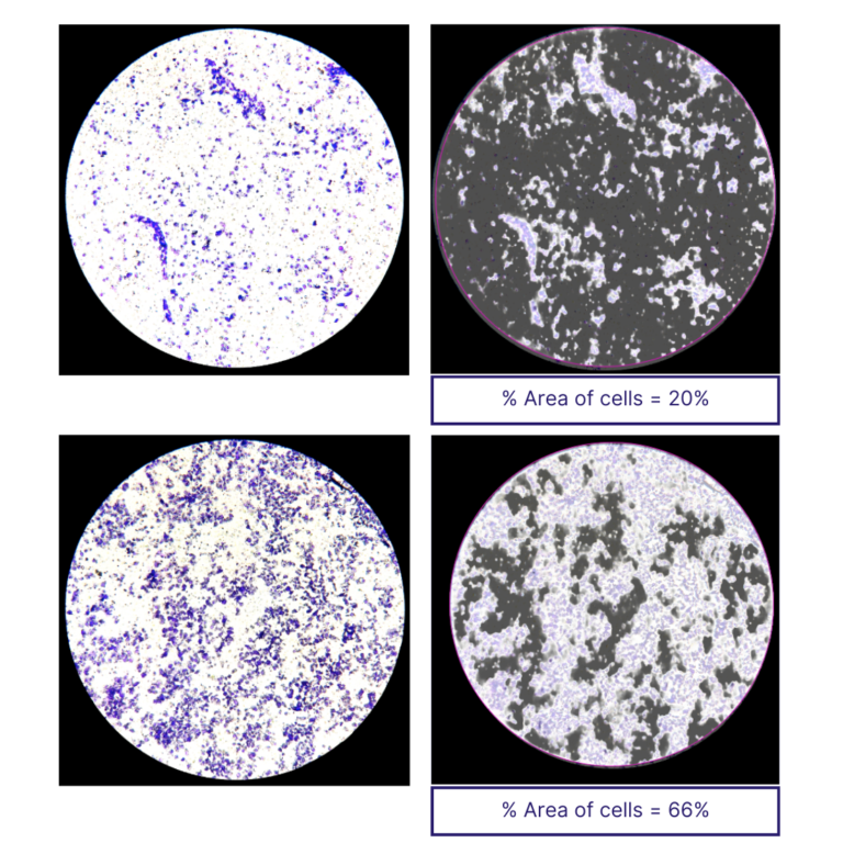
Looking for alternatives, Dr. Somasekharan’s team decided to test the SnapCyte™ technology to acquire and analyze their samples. The SnapCyte™ phone adapter dramatically improved the clarity and quality of cell images taken under the microscope, a process that was streamlined to just 15 minutes.
Moreover, the advanced SnapCyte™ AI algorithm was able to accurately detect the cells in the image after enhancing the staining contrast. The integration of SnapCyte™ into their workflow not only optimized their process but also ensured that the data collected was both reliable and accurate.
Key Benefits were:
Accuracy in Invasion Area Measurements: The AI-driven SnapCyte™ analysis provided the team with highly accurate data, essential for advancing their understanding of prostate cancer cell behaviors.
Time Efficiency:The complete imaging and analysis process was reduced to under 30 minutes, significantly optimizing protocol efficiency.
Environmental and Health Safety: SnapCyte™ eliminated the need for toxic dissolving agents and additional waste associated with other methodologies, offering a more sustainable solution.
Customer Testimony
“SnapCyte’s integration has significantly enhanced the precision and efficiency of our invasion assays, providing a solid answer to our previous data analysis challenge. We’ve shifted from spending hours on taking the images and figuring out various parameters on other platforms to analyzing our data within 30 minutes.”
“SnapCyte’s integration has significantly enhanced the precision and efficiency of our invasion assays, providing a solid answer to our previous data analysis challenge. We’ve shifted from spending hours on taking the images and figuring out various parameters on other platforms to analyzing our data within 30 minutes.”