Measure Cell Proliferation Using Confluency
Image-Based Cell Proliferation Assay
Cell proliferation plays a central role in cancer biology, drug screening, and regenerative research. However, traditional cell proliferation assay methods—like MTT, WST-1, or PrestoBlue—require destructive sampling and introduce variability across time points.
SnapCyte™ offers a smarter solution. By using cell confluency as a non-invasive proxy for proliferation, our image-based assay delivers accurate, reproducible results—without the need for dyes, extra reagents, or sacrificing any cells.
🔗 Want to learn more? Read our deep dive on cell confluency here.
How it works
How SnapCyte™ Proliferation Assay Work
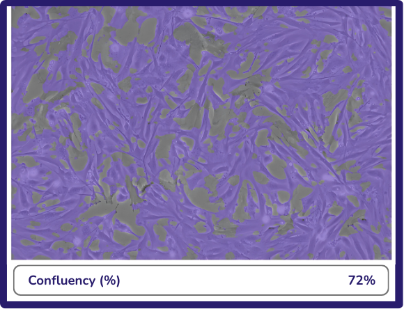
SnapCyte™ tracks cell proliferation by measuring confluency across time.
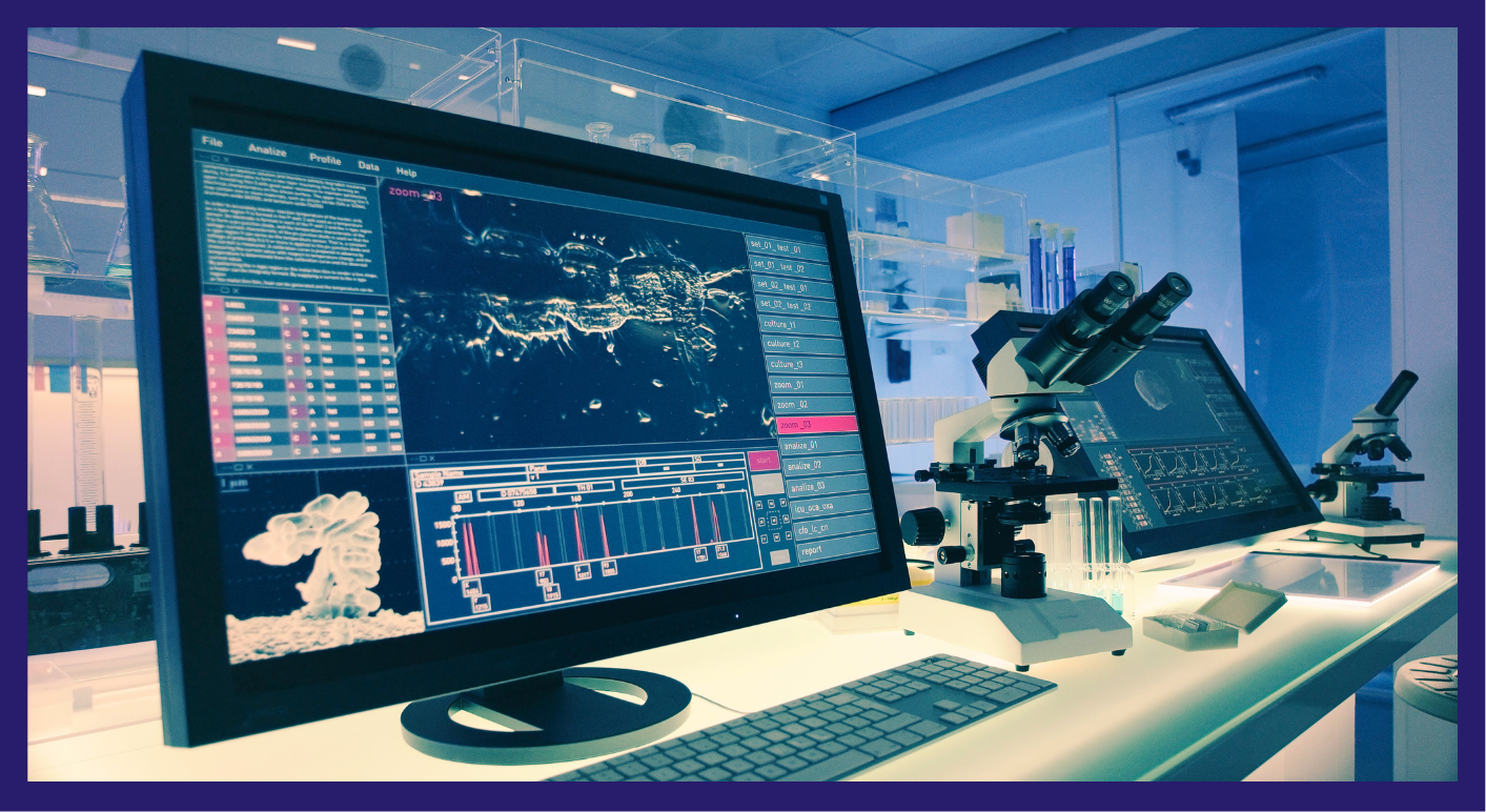
First, culture and treat your cells as usual. Then, capture brightfield or phase-contrast images using either a digital microscope or your smartphone with the SnapCyte™ adapter. Upload your images to the SnapCyte™ app, and our AI analyzes cell density changes over time.
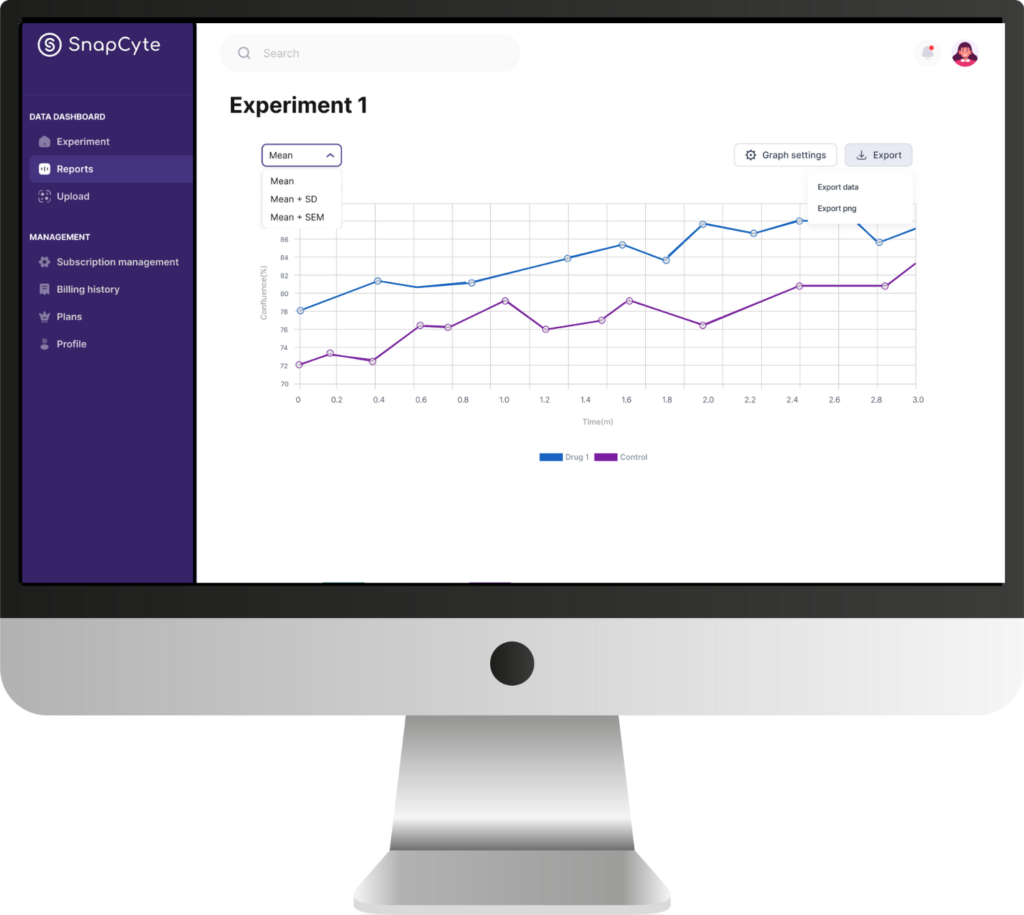
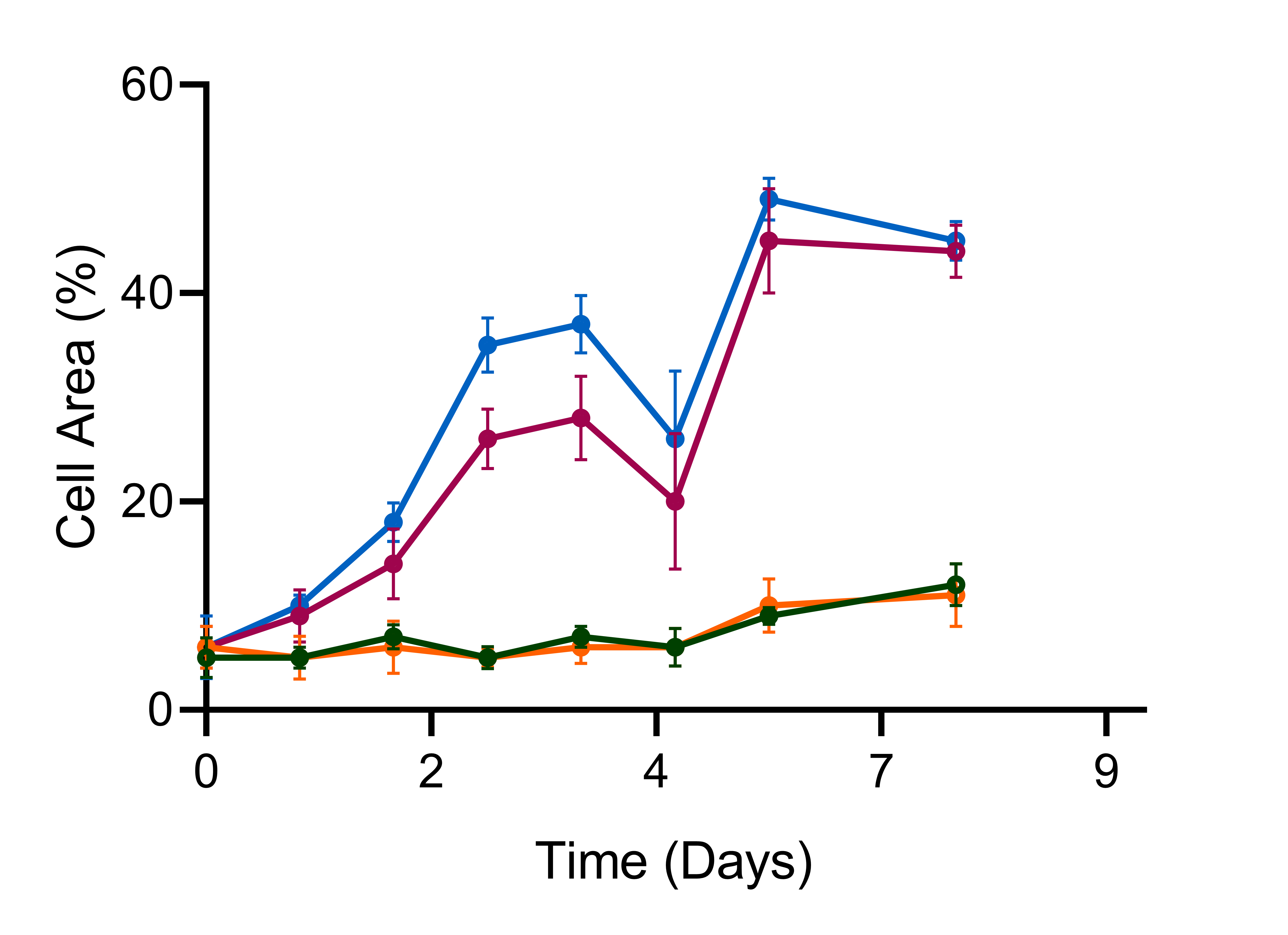
Because SnapCyte™ uses image-based measurements, it preserves your samples and generates a full proliferation curve from confluency data—all without disrupting the experiment.
📹 Scroll down to watch a short video on capturing images with your smartphone.
Why Use SnapCyte™ for Cell Proliferation?
Non-Invasive Monitoring
Track cell confluency over time to infer proliferation without dyes or cell death.Accurate and Reproducible
Our AI model applies standardized criteria across all time points and users.Save Time and Reagents
No need for reagents, destructive sampling, or multiple replicates per time point.Integrated Data & Images
SnapCyte™ keeps images, confluency plots, and growth curves together—ready for export or review.
Get Your Free Academic License
SnapCyte™ is free for academic use. Run a full cell proliferation assay using just images—no staining, extra reagents, or complex setups needed.
Case studies
Frequently asked questions
Most Frequent Questions and Answers
What types of cells can I use with SnapCyte™?
SnapCyte™ is compatible with a wide range of adherent cell types making it versatile for various research applications. You can also send sample images for analysis before starting your trial to contact@snapcyte.com.
How many images do I need from each well to obtain accurate, representative data?
The number of images needed per well depends on the format of the culture plate used and homogeneity of your culture. For example, in a 96-well plate, capturing 2 images at 10X magnification per well is typically sufficient for accurate data.
Do I need special equipment to capture images for SnapCyte™?
SnapCyte™ offers an adapter that allows you to acquire images using your smartphone and any basic microscope. It can also work with phase-contrast images taken with other microscopes, providing flexibility in how you capture and upload your data.
Can I track cell proliferation over time with SnapCyte™?
Yes, SnapCyte™ allows for continuous monitoring of cell proliferation by analyzing images captured at multiple time points, providing reliable data throughout your experiments.
How long does it take to capture and analyze images with SnapCyte™?
Analyzing each photo with SnapCyte™ takes less than 30 seconds. For example, if you’re doing an experiment in a 24-well plate, you need to take at least 3 images per well. So, it typically takes about 15 minutes to capture the whole plate.
Is there support available if I encounter issues using SnapCyte™?
Yes, SnapCyte™ offers comprehensive customer support, including detailed protocols and user guides in our help centre.