AI-Powered Cell Confluency and Proliferation Analysis in Apoptosis Research

Written by

Published on

Within the University of Leicester’s Department of Molecular and Cell Biology, a leading hub for innovative research, Dr. Joanna Fox’s laboratory is at the forefront of exploring the intricacies of cell death. Their work, particularly focused on the regulatory mechanisms of proteins like BAK within the intrinsic apoptosis pathway, is pivotal for advancements in understanding disease processes and therapeutic approaches. Click here for Dr. Fox’s publication list.

Challenges

A fundamental obstacle in the lab’s research was the need to increase data reproducibility. Their objectives centered on establishing consistent experimental starting points and accurately measuring cell proliferation in response to treatments that induce cell death. Traditional methodologies, including conventional cell counting for seeding and proliferation measurements, were found to be laborious and prone to variability, undermining the consistency and efficiency of their research efforts.

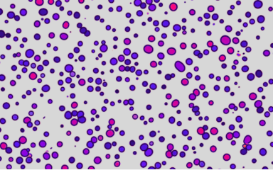
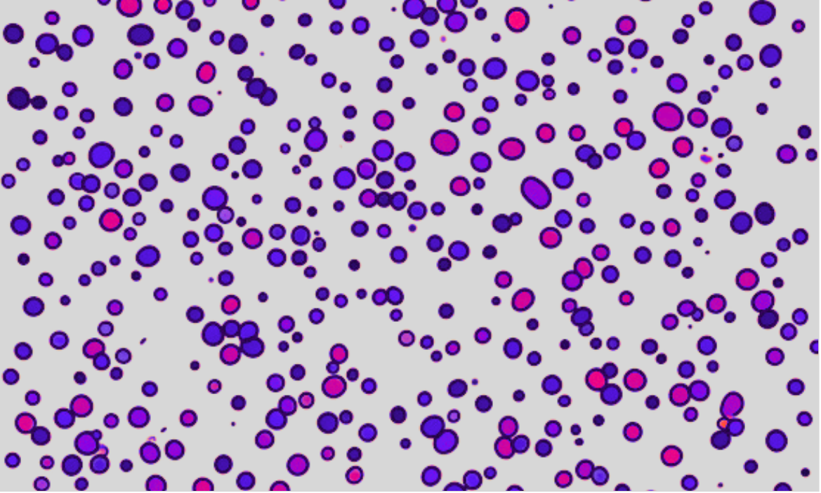
non-destructive cell proliferation assay using SnapCyte™
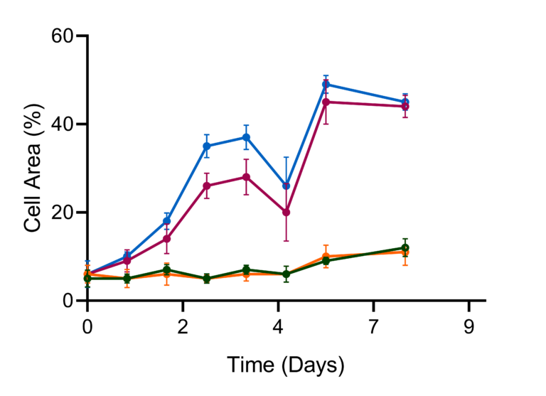
Figure 1. Senescence assay results for four treatment groups, confirmed using SnapCyte™ prior to staining with specific markers.

Key benefits of SnapCyte®

Looking for alternatives, Dr. Fox was introduced to SnapCyte at the 2023 EWCD conference in Italy. Recognizing its potential to increase their workflow efficiencies, she integrated the platform into her lab’s operations. With SnapCyte, they could achieve a high level of accuracy in measuring cell confluency and proliferation, streamlining their research process. The AI-based confluency and cell growth analysis provided by SnapCyte enabled precise assessment of seeding density and detailed evaluation of cell proliferation under different treatment conditions, leading to more reliable dataIn a senescence assay conducted in Dr. Fox’s lab, SnapCyte™ enabled the confirmation of treatment groups prior to the application of specific senescence markers, as illustrated in the Figure 1 depicting four treatment groups. This step was critical for verifying the effectiveness of treatments in inducing cell senescence before staining

“SnapCyte® has offered us rapid assessment of seeding uniformity and preliminary confirmation of cell death, thereby serving as a valuable quality control tool. Before progressing to more specific and costly reagents, SnapCyte® ensures the efficiency of our experiments and treatments.”

John Moss
PhD Student, Dr. Joanna Fox Laboratory, University of Leicester

In conclusion SnapCyte®:

  • Enhanced Data Reproducibility: By ensuring homogeneous seeding and standardizing the timing of downstream assays, SnapCyte increased the reliability of experimental data.
  • Improved Efficiency: The ability to quickly and accurately measure cell growth and responses to treatments saves valuable time and resources.
  • Cost-Effectiveness: SnapCyte’s pre-emptive verification of cell death before the deployment of costly markers and specific reagents underscored its economic benefits, reducing the overall expenditure on consumables.

Are you ready to try SnapCyte® toolbox in your lab?

SnapCyte™ is available at no cost for students and researchers. Instantly analyze cell confluency, proliferation, and viability—no plugins or coding required.

Resources

Related posts

Adherent Cell Count: More Than Just a Number

Cell Counting in Cell Culture: How to Reduce Variability

How Zymeworks Automated Spheroid Segmentation and Viability Readouts with SnapCyte