AI Image Analysis Toolbox for Cell Biology
Modern cell biology experiments generate large image datasets across multiple conditions and replicates. As experiments scale, interpretation becomes the bottleneck, not imaging. SnapCyte provides an AI image analysis toolbox designed to apply consistent analysis rules across cell biology assays, supporting confluency, cell counting, migration, and viability analysis.
Supported Assays
What Can You Analyze Today?
Adherent Cells
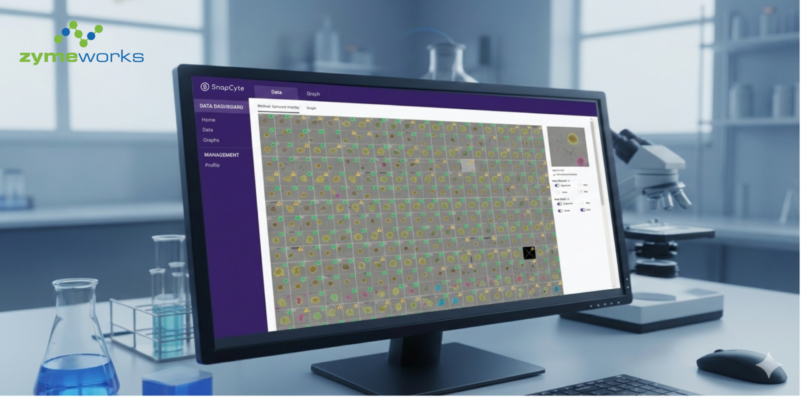
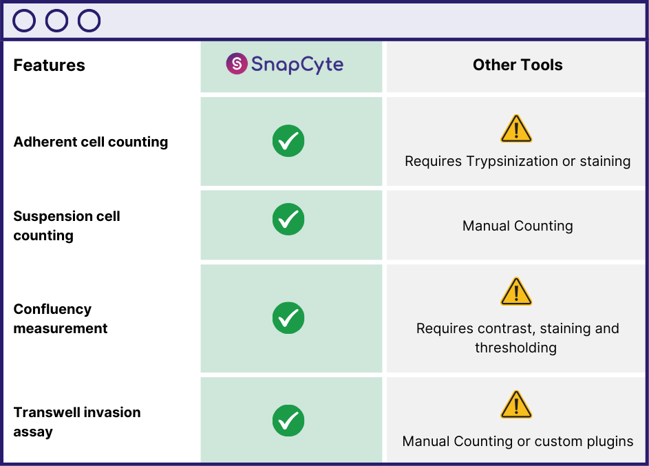
Count adherent cells directly in the well—no trypsinization or detachment needed. SnapCyte® analyzes brightfield images to deliver accurate, label-free counts. Ideal for preserving cells for downstream workflows like treatment, RNA extraction, or in vivo injection.
Suspension Cells
For single-cell suspensions (post-trypsinization or naturally suspended cultures), SnapCyte® supports automated counting and viability analysis using Trypan Blue. Just upload a hemocytometer image—SnapCyte® handles the rest.
Compatible with images form digital microscope or phone + adapter setups.
Measure confluency from brightfield with no staining required. Use it to calculate doubling time, standardize plating density, or ensure cells reach the right confluency before treatment.
With timepoint imaging, cell confluency and adherent count can be used to monitor growth curves for drug studies—offering a non-invasive way to track cell behavior over time without relying on absorbance or high-cost live-cell imaging systems.
Traditional invasion/transwell migration assays require manual counting or ImageJ-based analysis, which can be time-consuming and inconsistent. SnapCyte® automates this process by detecting and quantifying invaded cell areas as a percentage, giving researchers fast and objective results.
Coming soon!

How it Works
Three Steps to Results
Capture Your Image
Use a digital microscope or a smartphone with a microscope adapter to take high-quality images of your cells.
Upload & Set up Analysis
Upload your image to SnapCyte™ for instant AI analysis, or set up a timelapse experiment to track cell changes over time.
Get Results
Receive instant insights, including quantitative measurements, graphs and statistical error calculations, eliminating manual work.
Ready to try SnapCyte® AI in your lab?
Sign up for free academic access.
Why SnapCyte?
Designed for Real-World Lab Workflows.
SnapCyte™ was built by researchers who were tired of slow, inconsistent, and manual analysis workflows. Unlike ImageJ, which often requires macros, staining, or manual thresholding, SnapCyte™ automates your most common assays—cell counting, confluency, and invasion analysis—in a few clicks.
Whether you’re prepping a figure or validating growth before treatment, SnapCyte™ provides fast, reproducible results from standard brightfield or fluorescence images. No plugins. No coding. No waiting.
Need Help Using SnapCyte®?
Find answers, tutorials and trouble shooting guides in our help centre.
Frequently asked questions
How do I measure cell confluency with SnapCyte™?
SnapCyte™ automates cell confluency measurement using AI, providing accurate, reproducible results without manual thresholding. Simply upload your cell images, and SnapCyte™ will analyze confluency in seconds.
Can SnapCyte™ analyze invasion assays automatically?
Yes! SnapCyte™ quantifies the percentage of invaded cells in transwell invasion assays—eliminating the need for manual counting and analysis.
How does SnapCyte™ count cells in hemocytometer images?
SnapCyte™ detects individual cells from standard hemocytometer images. No manual counting required.
What type of microscope or camera do I need?
SnapCyte™ works with digital microscopes or smartphones with a microscope adapter. High-quality, well-focused images produce the best results.
Where can I find setup guides and troubleshooting help?
Visit our Help Centre for step-by-step guides on capturing images, setting up experiments, and optimizing analysis with SnapCyte™.
")