Summary
Cell confluency is a critical parameter in mammalian cell culture, and knowing the confluency level is important for controlling and optimizing cell growth, behaviour, and downstream applications. How is it defined, what affects it, and why is it so important to get just right? We explain all of that in this blog post. As datasets grow larger and more complex, consistent and objective analysis of cell confluency has become increasingly important.
What is cell confluency?
Cell confluency refers to the degree to which a culture of adherent cells in a dish or flask has filled a given area, typically measured as a percentage of the surface area covered by the cells. In other words, it’s the degree to which a culture dish or flask is “full” of cells. It is a very important parameter in cell culture and is used to determine when cells are ready to be subcultured, harvested, transfected, or otherwise used for experiments. The measurement of cell confluency is an essential step in maintaining healthy cell cultures and ensuring accurate experimental results.
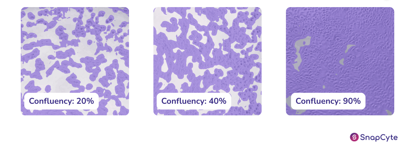
What do 50%, 80%, and 100% confluency mean?
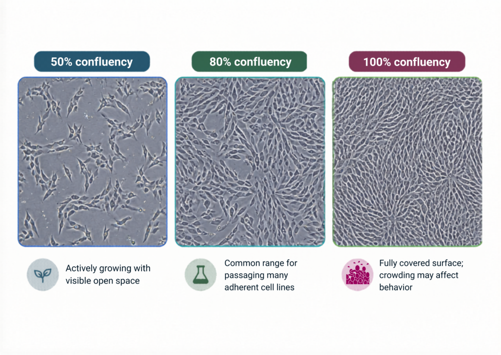
These three levels are useful checkpoints because they reflect different stages of adherent cell growth: early expansion, the usual passaging window, and full surface coverage.
50% confluency usually means the culture is still in an active growth phase. There is enough open space for cells to keep spreading and dividing, so this stage is useful for monitoring early proliferation.
70–80% confluency is often the practical decision point. Many adherent cell lines are passaged, harvested, or prepared for experiments around this range because the culture is dense enough to be useful but not yet overcrowded.
100% confluency means the culture surface is fully covered. This is also called a confluent culture. At this stage, cells may become tightly packed, slow growth, change behavior, or become stressed depending on the cell type.
These numbers are not universal rules. The ideal confluency depends on the cell line, morphology, protocol, and downstream application.
Why accurate cell confluency measurement matters
Confluency is an important consideration in cell culture as it can affect the growth and behavior of cells in culture. High confluency can lead to issues such as reduced proliferation, increased cell death, and changes in cell behaviour. Cell confluency can be controlled by adjusting the seeding density of cells, or by removing cells via subculture. This can affect the outcome of experiments and the quality of the cells being used for research, therapy, or other applications.
Why is cell confluency important?
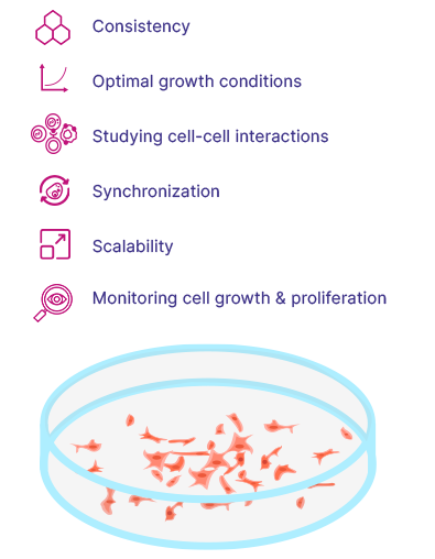
- Consistency: A confluent culture allows for a consistent and reproducible experiment, as all the cells are at the same stage of growth and division.
- Optimal growth conditions: Cells at confluency are typically grown under optimal conditions, which can minimize variability and improve the quality of the data generated from the experiment.
- Studying cell-cell interactions: Confluent cell cultures allow scientists to study the interactions between cells in a more natural and physiologically relevant manner, as the cells are in close proximity to one another and are able to interact through cell-cell junctions and signaling pathways.
- Synchronization: Confluent cell cultures can be used to synchronize cells for further experiments.
- Scalability: Confluent cell cultures can be used to scale up the production of cells for applications such as cell-based therapies and bio-manufacturing.
- Monitoring cell growth and proliferation: The percentage of confluency can be used as a measure of cell growth and proliferation, allowing scientists to monitor the growth of cells over time and make adjustments to the culture conditions as needed
Regularly measuring the confluency is also important because when cells are grown in culture, they are often used for downstream applications such as protein production, gene expression analysis, and cell-based assays. The confluency level can affect the outcome of these downstream applications, so knowing the confluency level allows researchers to control and optimize the conditions for these applications.
Additionally, in many cell culture systems, cell confluency is used as a parameter to determine when to split or pass the cells. The cells are usually split when they reach a certain confluency level, so that they can continue to grow and divide without becoming too crowded and overgrown. Therefore, knowing the confluency level is essential for maintaining the health and vitality of the cells in culture.
Why visual confluency estimation becomes subjective
In practice, cell confluency is rarely assessed from a single image or condition. Large experiments often include multiple replicates, timepoints, and treatment groups, making consistent interpretation difficult. Visual estimation and manual thresholds can introduce variability, especially when differences between conditions are subtle.
How to measure cell confluency in cell culture
There are several methods for measuring cell confluency, each with their own advantages and disadvantages. For a comparison of measuring methods, check out our new eBook: How to Measure Cell Confluency.
Manual visual estimation of cell confluency (aka. 'Guesstimation')
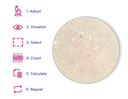
One common method is manually estimating cell confluency using a microscope and visually inspecting the cell culture (sometimes referred to as ‘guesstimation’ due to the not so accurate data outcome). This method involves estimating the area of a cell culture flask or dish covered by cells and comparing it to the total surface area, resulting in a confluency percentage. The disadvantage of this method is that it is time-consuming and subject to observer bias. In many workflows, confluency estimation is closely tied to cell counting methods used to assess growth and viability.
One common method is manually estimating cell confluency using a microscope and visually inspecting the cell culture (sometimes referred to as ‘guesstimation’ due to the not so accurate data outcome). This method involves estimating the area of a cell culture flask or dish covered by cells and comparing it to the total surface area, resulting in a confluency percentage. The disadvantage of this method is that it is time-consuming and subject to observer bias. In many workflows, confluency estimation is closely tied to cell counting methods used to assess growth and viability.
Colorimetric assays for estimating cell growth
A second method for measuring cell confluency is the use of cell-specific dyes or stains. These dyes or stains bind specifically to certain cell components, such as nuclei, and can be used to visualize the cells in a culture. By analyzing the intensity of the dye or stain, it is possible to estimate the number of cells in a culture. Although this method is more accurate than visual estimation or image analysis, it is also more expensive and time-consuming.
Image-based cell confluency analysis
A number of tools offer image-based confluency measurements. These either consist of a software package that analyzes images taken from a microscope, or are integrated instruments that image and analyze your cell culture directly.
Digital image analysis software can be used to automatically quantify cell confluency from microscope images. These software programs can recognize cells and calculate the ratio of the cell-covered area to the total area, providing a more objective measurement than manual estimation. This method is more objective than manual counting, but it can be affected by variations in cell size, shape, and colour.
AI-based cell confluency software
The most recent development in cell confluency tools is the incorporation of AI-based algorithms to analyze cell culture images. These tools apply the same analysis rules across all images, helping reduce variability caused by manual interpretation. They require no manual input and are a useful way to train students when initially wanting to learn to visually estimate a cell culture’s growth rate and overall health.
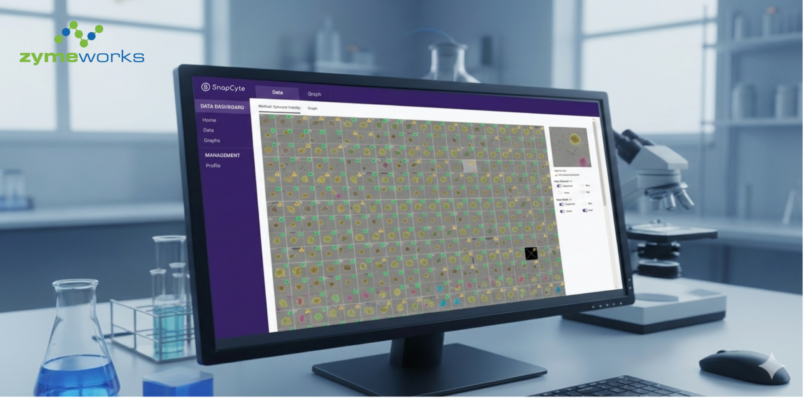
SnapCyte® is one example of an AI-based platform designed to support objective confluency analysis directly from microscopy images, without staining or specialized hardware.
Manual vs AI-based confluency measurement
Manual / Visual Estimation
- Subjective and user-dependent
- Limited reproducibility across users and timepoints
- Not scalable to large datasets or multiple experimental conditions
- Fast for quick culture health checks
AI-Based Confluency Analysis
- Applies the same analysis rules across all images and conditions
- Reduced user-dependent bias and variability
- Handles morphological and imaging variability
- Scalable to large, high-throughput datasets
Measure Cell Confluency More Consistently
Use SnapCyte® as cell confluency analysis software to measure confluency from brightfield microscopy images and reduce visual estimation bias across users, timepoints, and experiments.

Janny Marie L. Peterslund, PhD
I am a biologist and science communicator with experience in stem cell biology, biotech validation, and technology commercialization. I focus on turning complex scientific concepts into clear, useful content for researchers and technical audiences.

")
")
")