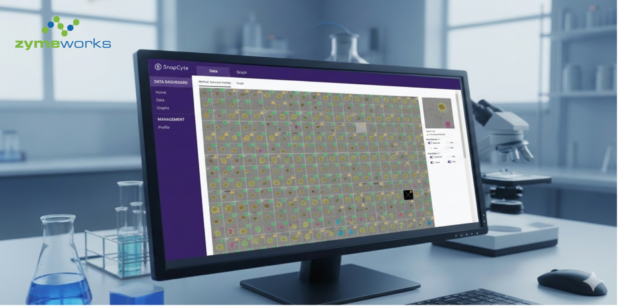
Zymeworks’ antibody-drug conjugate (ADC) team turned to AI to address high-throughput challenges associated with their workflow for spheroid segmentation and assay quality control (QC) of a large number of spheroid images. By working with SnapCyte, they replaced hours of manual review with fast, reproducible and consistent image-based metrics. To understand how this transition happened, we spoke with the team about the issues they faced and what changed after adopting a custom SnapCyte AI model.
Tell us a little about the project and the problem you were facing.
“We run large-scale spheroid viability assays as part of our screening pipeline. Each plate contains multiple wells imaged across brightfield and fluorescent channels, and our downstream viability metrics depend heavily on accurate segmentation of each spheroid.
The challenge was that the analysis module built into our imaging system didn’t generalize well to real experimental variation. When spheroids were perfectly circular and well-centered, the software performed reasonably. But in many plates, spheroids were irregular, shifted, dim, or partially fragmented, and that’s where the segmentation routinely failed. Because of this, every plate could required up to 1 hour of manual QC, reviewing each well individually to check for mis-segmented objects, tracking down outliers, and determining whether the viability value reflected biology or simply a segmentation error, leading to data exclusion of wells and higher assay variability.”
“We tried obvious fixes like adjusting thresholds and analysis parameters, but those only solved a few isolated cases. The core problem was that the workflow couldn’t adapt to the full range of morphological variations seen across plates and cell types. For a high-throughput assay like this, that type of manual burden wasn’t sustainable.”
Why did you decide to partner with SnapCyte?
“What stood out was SnapCyte’s ability to build a custom model trained on our exact assay format. They didn’t offer a generic solution. They studied our failure cases, reviewed our analysis criteria, and benchmarked directly against our internal analysis pipeline. That gave us confidence that the final model would actually solve the problems we were seeing, not just improve them slightly.”
How did SnapCyte solve the problem?
“SnapCyte developed a multi-channel AI segmentation model trained on our brightfield and fluorescent channel. The model handled:
Precise spheroid boundary segmentation across diverse morphologies
Automated debris exclusion
Fluorescence quantification aligned with our normalization method
QC flagging so we only review wells that truly need attention
Once deployed on the SnapCyte® platform, the workflow became extremely simple: upload a plate image, run analysis, review flagged wells, export results.”
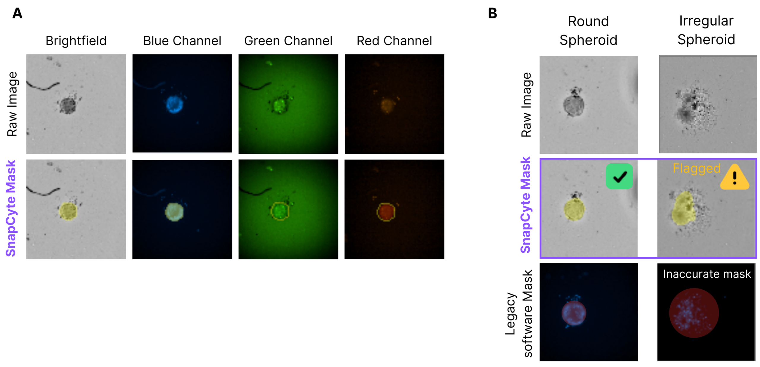
A) Brightfield and fluorescent with corresponding SnapCyte masks, showing consistent spheroid segmentation across imaging modalities. B) SnapCyte accurately segments both round and irregular spheroids, while irregular cases are automatically flagged for review. In contrast, legacy software produces blue pixel-dependent masks that fail to adapt to spheroid morphology.
What was the biggest benefit of using SnapCyte?
“The time savings were also significant. What used to take up to 1 hour of manual QC for large assays now takes less than 5 minutes, and we only look at the small number of wells flagged by the model instead of scanning every well manually. Overall, the workflow became faster, more reproducible, and far more scalable.”
How would you describe working with SnapCyte and developing a custom AI model from start to finish?
“Working with SnapCyte was straightforward and efficient. The process began with us providing the raw data along with examples of problematic cases where our previous analysis pipeline was failing. From there, SnapCyte handled the full development workflow — including data annotation, defining the analysis criteria, and establishing the readouts. They delivered the first working model quickly.
We went through iterative cycles where we supplied additional images and edge cases, and SnapCyte refined the model based on our feedback. Our involvement remained light throughout the process; most of our work consisted of reviewing outputs, giving comments, and validating updates. Communication was clear, and we always knew what was needed on our end.
Once the model reached the performance level we required, SnapCyte deployed it on the SnapCyte® platform, giving us a streamlined workflow for full-plate analysis, QC flagging, and results export. Overall, the process from start to finish required minimal effort from our team and resulted in a reliable, scalable tool that now supports our high-throughput assays.”
In conclusion SnapCyte enabled Zymeworks' ADC team to reduce analysis time by 92%, optimize the workflow and improve their QC process significantly.
Interested in starting a custom AI image analysis project? Learn more about how we work here.

")
")
")