Getting started with SnapCyte®
SnapCyte® is AI-powered cell image analysis. No coding, no manual counting, no staining required for most assays. This guide takes you from sign-up to your first result.
1. Create your account
Sign up with your institutional email at app.snapcyte.com/signup. Verify your email and you’re in.
2. Choose how you'll capture images
Digital microscope
Capture your images and export them as TIFF, JPEG or PNG. Upload your images at app.snapcyte.com. Best for labs with dedicated digital microscopes or camera attachments. No extra setup.
SnapCyte Lens® + adapter
A free app that lets you capture images from your phone and syncs them automatically to your experiments. Hold your phone to any standard upright microscope eyepiece, or use a phone adapter for better stability.
Don’t have an adapter? We recommend Celestron NexYZ adapter and Swift 5.0 Megapixel Digital Camera.
Either path produces the same results. The difference is in image quality, existing set up in your lab and convenience.
3. Pick your assay and run your first experiment
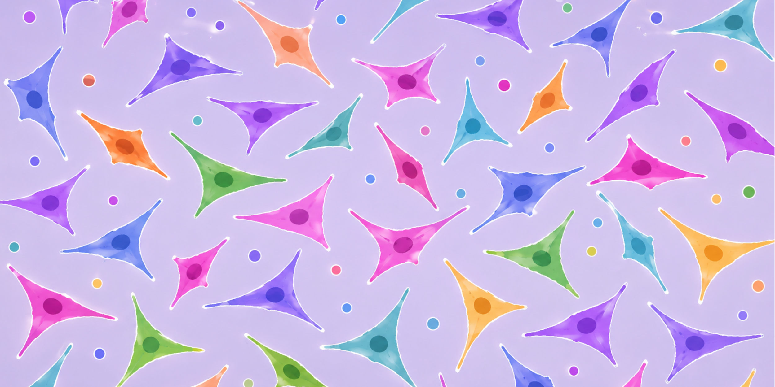
Cell Growth
Confluency percentage and adherent cell count from a single brightfield image. Best for tracking how cells grow over time, measuring drug effects on proliferation, or checking size changes in cells.
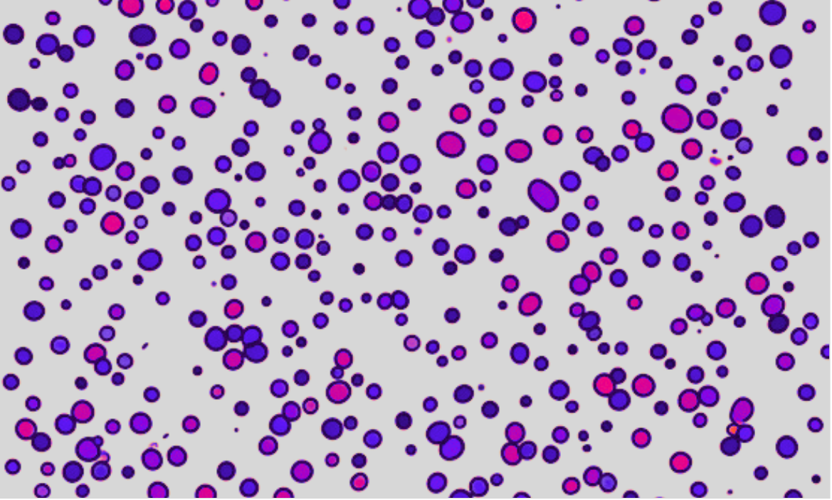
Hemocytometer Count
Total cell count, live/dead viability, and cells/mL from suspension. Works with Trypan Blue or without.
Invasion Assay
Quantifies the percentage of membrane area covered by crystal violet-stained, invaded cells from transwell assays.
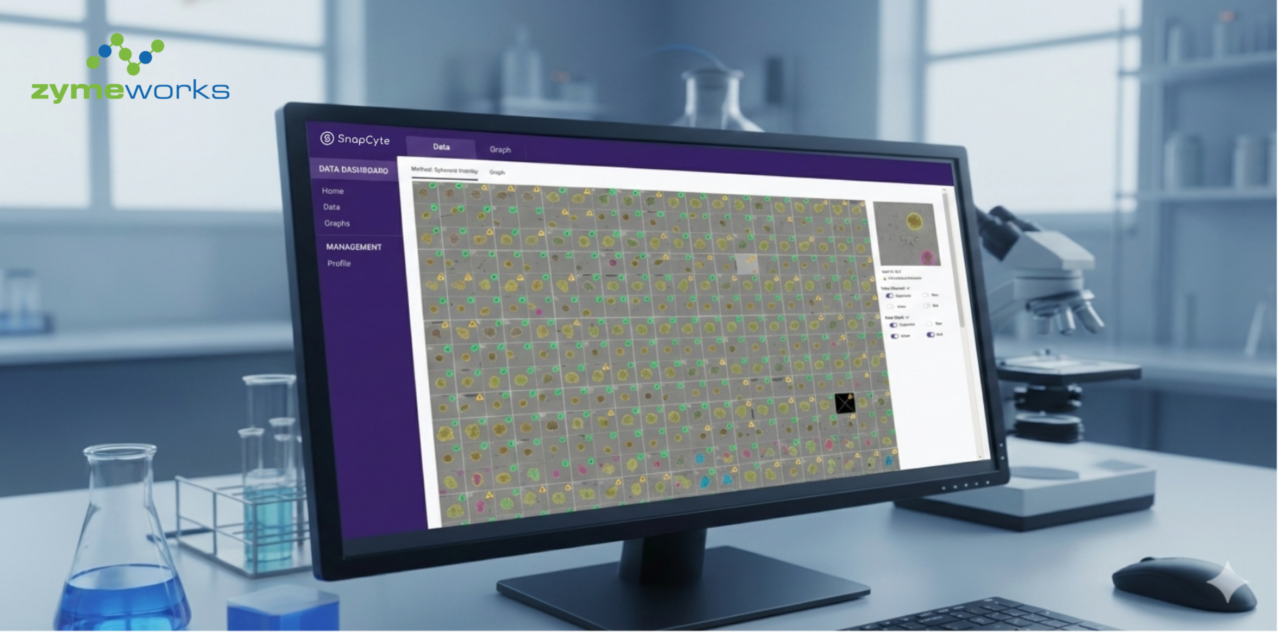
Create a new experiment, upload images, organize them into groups (your conditions) and time points (if needed). SnapCyte returns per-image results plus mean and SD across each group.
ROI: We automatically detect the cell region and suggest an ROI if you have a lot of dark background. Always check the results to make sure empty regions, black border, or off-target area are excluded by an ROI. If not, they will be counted as total surface area and will make your confluency or stained cell % read artificially low.
4. Export and use your data
Three options when you’re done:
- Export data : Full Excel file with your group structure, per-image results, and group means with SD
- Export graph : Bar graph (single time point/plate) or line graph (multi time points/plates), ready to share
- Export images: Download all images from the experiment as a bundle
Frequently asked questions
My upload is stuck
Check file size, single batches are capped at 200 MB and it’s best to make sure each image is not larger than 10 MB. Split larger experiments into multiple batches.
Analysis failed
Most often caused by >2000 cells in one image or the wrong file type. Retry the analysis and check your image has the right format.
I did't get the verification email
Some academic institutions block unfamiliar email domains. Try again and if you were not able to get the email try another email address with a different domain.
I forgot my password
Use Reset Password on the login screen. The link expires in 30 minutes.
Do credits roll over?
Subscription limits reset monthly. Top-up credits never expire.
")