Transwell Migratio/Invasion Assays
AI Image Analysis: Invasion & Migration Assay
Invasion and migration assays are essential for studying cancer metastasis, immune response, and cellular motility. Yet, traditional approaches—like optical density (OD) readings or manual counting—often lead to inconsistent and labor-intensive workflows. OD-based methods are sensitive to staining variability, and manual counting is subjective and time-consuming.
SnapCyte™ offers a better approach. Our AI-powered platform analyzes stained images of migrated or invaded cells, providing stained-area-based quantification that improves both reproducibility and accuracy—no manual counting required. Read our complete breakdown of Transwell migration and invasion assays to better understand the workflow and key differences.
How it works
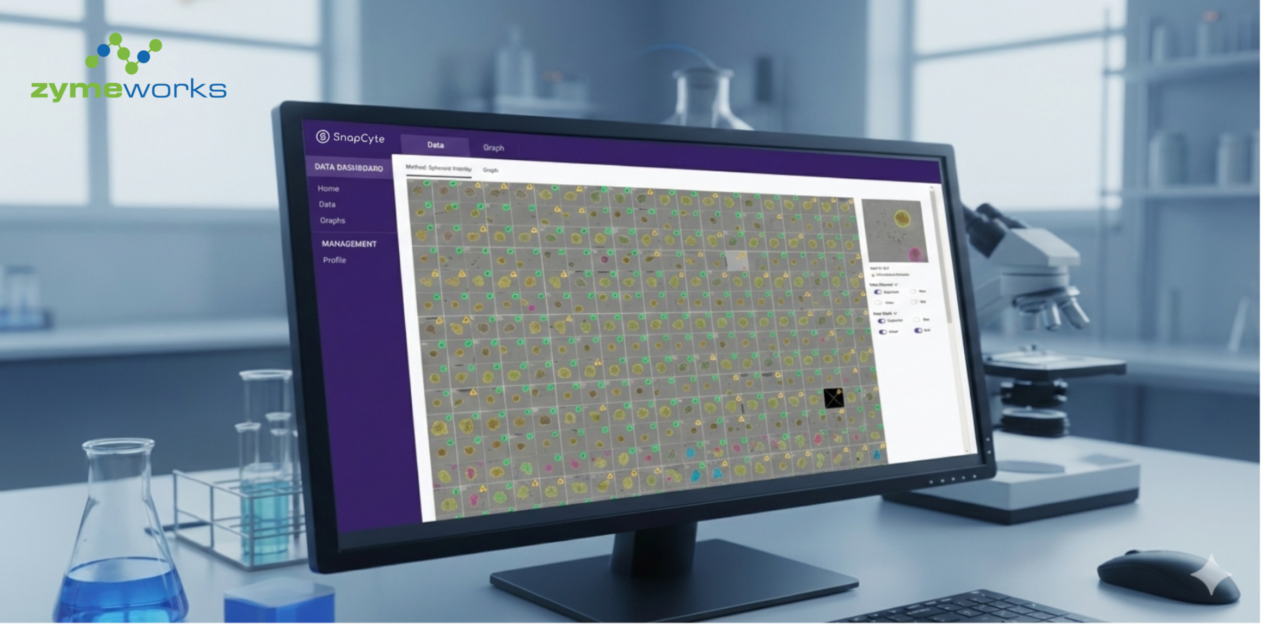
How SnapCyte™ Invasion Assay Work
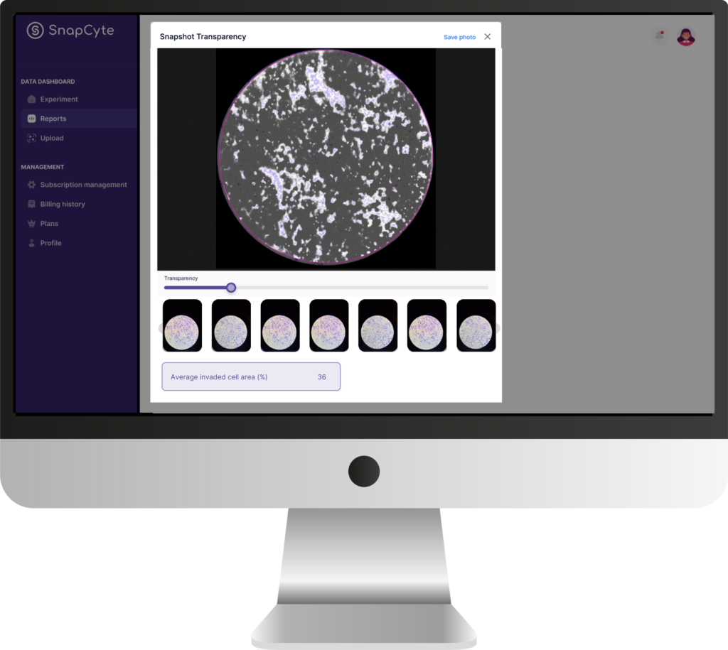
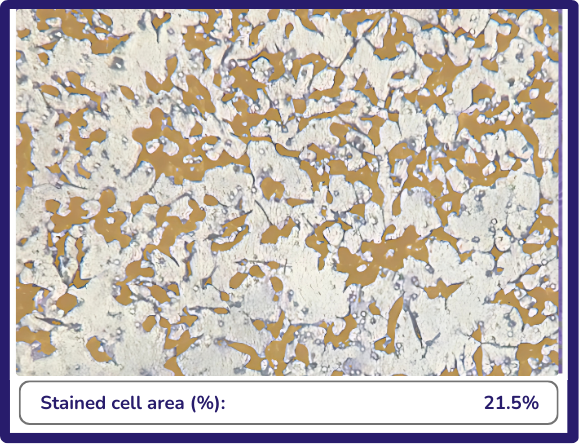
After staining migrated or invaded cells on the transwell membrane, capture multiple 10X images per well using your microscope or smartphone adapter. Upload the images to SnapCyte™, and the AI quantifies the percent area covered by stained cells, offering a fast and objective readout for both migration and invasion assays.
SnapCyte™ removes the need for manual counting, ensuring consistency even when cell boundaries are unclear or stain intensity varies.
📹 Scroll down to see a short video on how to image and analyze your migration and invasion assays with SnapCyte™.
Case studies
SnapCyte™ integration has significantly enhanced the precision and efficiency of our invasion assays, providing a solid answer compared to our previous data analysis challenge.

Why Use AI Image Analysis for Migration and Invasion Assays?
Automatically measures stained area—no counting or threshold tuning required.
Analyze each image in under 30 seconds, eliminating tedious manual steps.
Consistent analysis logic minimizes user variability and improves data quality.
SnapCyte™ stores your images, overlays, and metrics for seamless tracking and reporting.
Works with both migration (uncoated inserts) and invasion (matrix-coated inserts).
Get Your Free Academic License
Whether you’re performing a migration or invasion assay, you can now automate your analysis and eliminate manual error.
Frequently asked questions
Most Frequent Questions and Answers
How does SnapCyte™ improve the accuracy of invasion assays?
SnapCyte™ enhances accuracy by providing an stained-area based assessment of cell invasion, reducing the subjectivity and variability associated with manual counting.
Can I use SnapCyte™ with any type of transwell insert?
Yes, SnapCyte™ is compatible with a variety of transwell inserts, making it adaptable to different experimental setups.
What magnification is recommended for capturing images?
You can use 4X or 10X magnifications to acquire images. However, SnapCyte™ is optimized for 10X magnification, which balances detail and field of view for accurate analysis.
How long does it take to analyze the images with SnapCyte™?
SnapCyte™ processes each image in under 30 seconds, providing rapid results without compromising accuracy. Taking pictures of a 24-well plate would take around 15-20 minutes.
What kind of data output can I expect from SnapCyte™?
SnapCyte™ provides detailed quantitative data, including the percentage area of migratory (stained) cells and graphical representations of your results with various statistical analysis, ready for publication or further analysis.
")